DIY Brains!
 Well, not quite. But they are vaguely brain-like blobs…
Subject areas: Neuroscience, Developmental Biology
Vocabulary:
organoid – a mass of tissue that physically resembles and may have some physiological properties of a natural organ in an animal’s body, but is incomplete in form and function. They may be naturally occurring, such as some kinds of tumors, or they may be artificially created, as demonstrated in the paper discussed here.
pluripotency and stem cells have been discussed in previous posts – in short, stem cells are pluripotent, meaning that they can make several different cell types when they proliferate and differentiate. Most adult cells are not pluripotent and can only make more of the same cell type.
The article:
Lancaster, M.A. et al. Cerebral organoids model human brain development and microcephaly. Nature 501:373-379, 2013. doi:10.1038/nature12517
Background
It is clear that the pluripotent stem cells of the early embryo are able to form all of the many different cell, tissue, and organ types in the an animal or human being. It is equally well understood that correct functional formation of these tissues and organs requires precisely timed and placed chemical signals that can induce the patterns of differentiation needed to make the cells in one area produce heart muscle while the cells in another area produce neurons in the brain. However, it is not known just how much can cells do on their own, in the absence of the usual chemical organizing signals?
Although certainly an interesting question in itself, the impetus for this research was actually more medically related. The group wanted to understand the biology that leads to microcephaly, a developmental defect that causes an abnormally small head and brain. To that end, they thought to take normal and microcephalic pluripotent stem cells, and examine how they would behave in a three-dimensional culture system.
What they Did
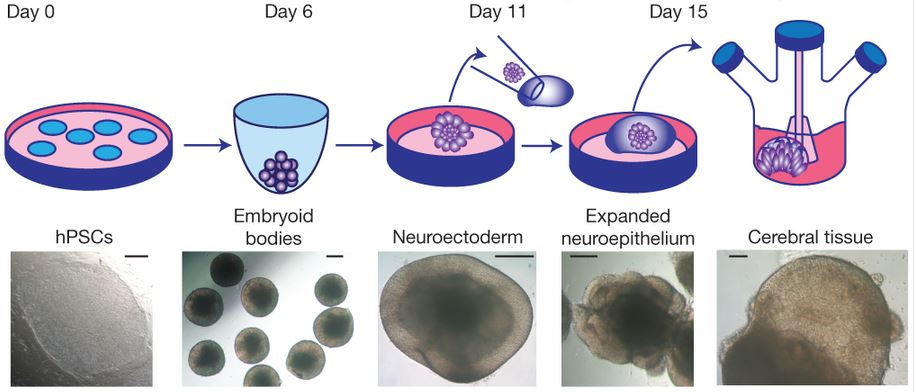
The diagram above is almost self-explanatory. First the human pluripotent stem cells are cultured in a liquid suspension until they formed small masses called embryoid bodies. At this point, the cells have not differentiated much yet, but by changing the growth medium, they could cause differentiation so that layers form, including an outer neuroectoderm layer (which is a precursor to brain tissue). This is then cultured in a gel suspension so that it can form in three dimensions (as opposed to flattening to the base of a culture dish), and you can see further differentiation into neuroepithelium as well as morphological changes that are reminiscent of actual cerebral brain tissue, even to the point of having a fluid-filled cavity (like ventricles in the brain that are filled with cerebrospinal fluid).

This cerebral organoid has retinal tissue (black arrow). Scale bar is 0.2mm
You can see in the photo to the right that an eye-like protrusion has formed. This is further evidence that brain-like structures are forming because the retina actually forms from the same precursor tissue as the brain. They also stained the organoid for neural-specific markers to confirm that it is actually neural tissue and not just something that has the same shape as a developing brain. Using different markers, they were actually able to show not just that neural tissue had arisen, but that the organoid stained for several different brain regions (see below), and that they were organized separately, not just mixed cell types differentiating randomly. Of course, closer examination showed that the differentiation and organization was not perfectly like that in a developing embryonic brain, but it does appear to have many of the general characteristics that could make this a reasonable model system for studying early brain development.
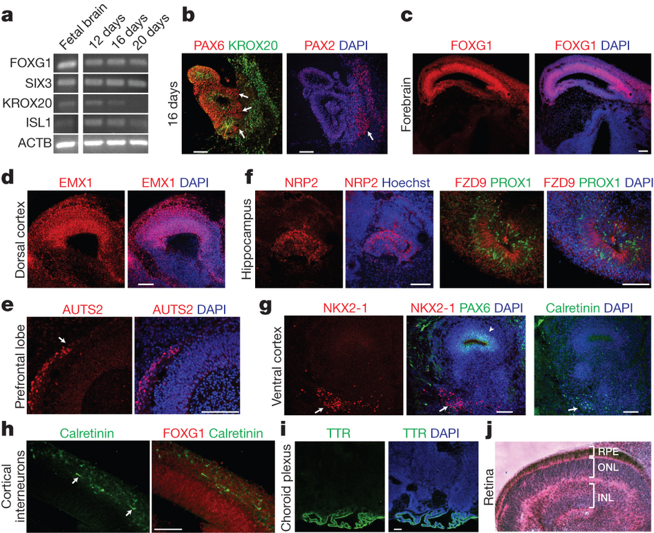
Click for full picture. Stains for the different areas are labeled, e.g. forebrain, hippocampus, etc. DAPI is a stain that shows the cell nuclei.
Not only does the overall organoid have different regions that correspond to brain parts, the development of the brain in layers is also somewhat preserved in this organoid culture system. The group even considered whether human-specific features were maintained in this system, so they compared the creation of organoids from human stem cells and from mouse stem cells. They were able to confirm that not only was the organoid larger for humans, but the regions within the organoid that should be thicker or larger in humans than in mice were in fact so.
Now what about microcephaly. After all, that was the point of trying to create this culture system. Could microcephaly be modeled in this system? So, they collected skin fibroblasts from a patient with severe microcephaly, and treated them by standard protocols to turn them into pluripotent stem cells. They then cultured these stem cells as they did before to make the brain organoid. The figure below shows quite clearly that there are significant differences in the cerebral organoids made from microcephalic patients in comparison to those from healthy controls. Microcephalic organoids are not just smaller, they also exhibit poor or aberrant organization. There also seem to be fewer and smaller proliferation zones (which is where brain cells are “born”).
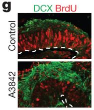
We can see changes between cerebral organoid from normal stem cells (top) and microcephalic stem cells (bottom). DCX indicates a neuronal differentiation, while BrdU indicates proliferating cells. There is clear disruption to the normal patterning.
This particular patient had mutations in a gene called CDK5RAP2 leading to production of truncated and presumably non-functional protein. This gene product is normally found in the centrosome, where it likely plays a role in centriole and mitotic spindle formation, and the Golgi complex, where its role is unclear. Mutations in CDK5RAP have been linked to some other cases of microcephaly. So, the researchers did some experiments to see if they could “rescue” microcephalic organoids by inserting genes for normal CDK5RAP and make them more healthy. The results were equivocal, partly because it was complicated by the fact that too much CDK5RAP was toxic. However, they were at least able to show that if they inserted something to knock down CDK5RAP in normal cells, it caused disruptions somewhat similar to the microcephalic organoids.
What they learned
First, they learned that they could make something vaguely brainlike out of stem cells. That is pretty cool in itself. The neuroepithelium has layers like a real developing brain, and there is compartmentalization of various functional markers. It even started to make part of an eye! They also found that making this cerebral organoid from cells taken from microcephaly patients led to malformations, suggesting that it could be used as a model for investigating what is happening in microcephalic embryos at the tissue and cell level. Finally, they have further evidence that CDK5RAP is involved in proper formation of the early brain.
No comments
Be the first one to leave a comment.